
HEART-GeN Research Program
Research Vision
The HEART-GeN lab studies how human genetic variation influences RNA regulation and cellular states across brain development, aging, and disease, and which of these mechanisms can be experimentally tested. We integrate human genetics, transcriptomics, epigenomics, statistical modeling, and experimental systems to move from genetic variation to regulatory mechanisms, cellular consequences, and disease-relevant biology.
Our work is organized around three connected ideas:
- Regulatory mechanisms. Genetic ancestry, RNA splicing, isoform choice, gene expression, and DNA methylation are the molecular layers through which DNA variation acts in the brain. Rather than relying on gene-level expression alone, we ask how genetic effects operate through transcript choice, epigenetic state, cell-type-specific regulatory programs, and developmental transitions.
- Biological consequences. We study these mechanisms where they matter: cortical development and neuronal maturation, brain aging, schizophrenia, and Alzheimer’s disease and related dementias.
- Mechanistic validation and translation. Interpretable genomic AI, human brain tissue, cerebral organoids, iPSC-derived models, and perturbation studies let us test which regulatory events are causal, and move toward ancestry-aware precision neuroscience.
Three principles connect this work:
- Mechanism over association. We prioritize the regulatory events and cellular states through which genetic variation may influence brain function and disease, and we distinguish predictive molecular signatures from mechanisms supported by genetic, longitudinal, or experimental evidence.
- Generalizability by design. We investigate ancestry-associated and context-dependent effects, evaluate whether findings transfer across cohorts, and build tools that make genomic analyses more inclusive. Genetic ancestry is one dimension of human genomic variation – not a biological proxy for race, and not a complete explanation for health disparities. Social, environmental, and structural factors also shape brain health and access to care.
- Computation linked to biology. We pair interpretable statistical and machine-learning methods with postmortem human brain datasets, single-cell and spatial genomics, cerebral organoids, and iPSC-derived models, so that computational predictions become testable hypotheses.
Where we are going. Our established programs in ancestry-aware regulatory genomics, RNA isoform regulation, schizophrenia, and brain aging give us a foundation for three long-term objectives: an experimentally validated atlas of regulatory programs across brain development, aging, and ancestries; molecular signatures of regulatory dysregulation that could support patient stratification; and a pipeline that nominates noncoding variants and RNA processing events as candidate therapeutic targets. These are goals we are working toward, and we pursue them through open resources and by training a diverse workforce fluent in both computation and experiment.
Ancestry-Aware Regulatory Genomics
How does human genetic diversity shape gene regulation across brain regions, cell types, and the lifespan?
Individuals with recent African ancestry account for less than 5% of large-scale genomic research cohorts (Gurdasani et al. 2019; Sirugo et al. 2019), yet Black Americans are roughly 20% more likely to experience serious mental health problems than the general population (U.S. HHS Office of Minority Health), and genetic differences have been linked to divergent antipsychotic responses across populations (Murphy and McMahon 2013). These statistics describe socially defined groups and reflect structural, environmental, and clinical factors as well as biology. Our question is narrower and mechanistic: which regulatory effects in the human brain differ with genetic ancestry, and do those effects replicate across cohorts?
We integrate global and local ancestry, expression quantitative trait loci (eQTLs), allele-specific expression, and transcript-level measurements from admixed and diverse cohorts. A major aim is to distinguish reproducible genetic mechanisms from cohort-specific or technical effects, improving the interpretability and portability of genomic discoveries.
What we have found. In the largest analysis of its kind, we profiled gene expression across four brain regions (caudate nucleus, dorsolateral prefrontal cortex, hippocampus, and dentate gyrus) in neurotypical Black Americans. Thousands of genes showed ancestry-associated expression, much of it explained by local genetic variation, with immune and vascular pathways among the most enriched. This work established that ancestry-aware analyses are both feasible and informative in the human brain, and it motivates our ongoing work on aging and neurodegeneration.

Dr. Benjamin discussed this work on NPR:
Representative publications
- Benjamin KJM, et al. Analysis of gene expression in the postmortem brain of neurotypical Black Americans reveals contributions of genetic ancestry. Nature Neuroscience (2024). Article
- Benjamin KJM. RFMix-reader: Accelerated reading and processing for local ancestry studies. bioRxiv (2024). Preprint
RNA Processing and Neuronal Maturation
When does transcript choice change cellular function rather than simply mark a cell state?
Most genes produce multiple RNA isoforms through alternative splicing and transcript usage, and a gene’s total abundance can stay constant while the cell switches which transcript it makes. We investigate how coordinated isoform transitions accompany cortical development and aging, whether specific transitions regulate neuronal maturation, and how disease-associated genetic variants perturb these programs.
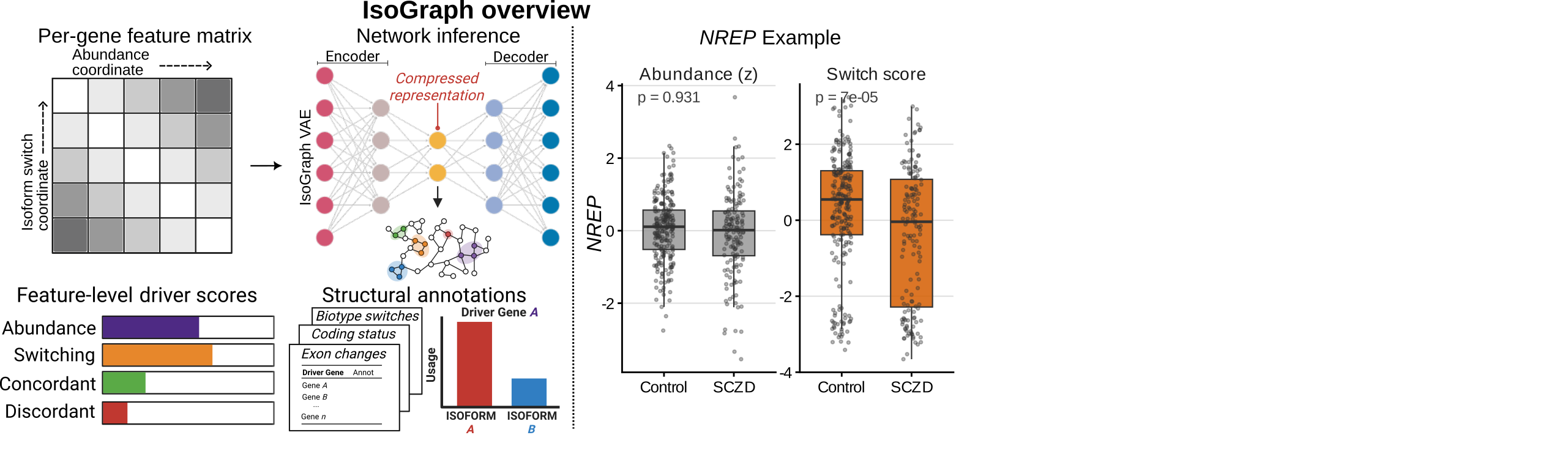
Our approach combines human brain transcriptomics, statistical genetics, and isoform-aware network methods. Because transcript switching is invisible to gene-level analyses, we developed IsoGraph, a multiplex network framework that jointly models isoform choice and gene abundance to identify coordinated transcript-switching programs. We then connect candidate programs to experimental models, with a focus on cortical projection-neuron maturation.
What we have found. Applying IsoGraph to the aging human brain, we resolved programs of coordinated transcript choice that are distinct from gene-abundance programs, including isoform switches associated with aging that would be missed by conventional differential expression.
Representative publications
- Bennett, Johnson, Benjamin KJM. IsoGraph resolves coordinated transcript choice and gene abundance in the aging human brain. bioRxiv (2026). Preprint
Schizophrenia and Neurodevelopment
How do genetic and molecular changes alter the development and function of disease-relevant neural cell populations?
Schizophrenia is a neurodevelopmental disorder with substantial genetic risk, yet most risk variants are noncoding and act through gene regulation. We study schizophrenia across postmortem human brain regions – the caudate nucleus, dorsolateral prefrontal cortex, and hippocampus – and in experimental models of human brain development. Our work addresses cell-type-specific regulation, sex-associated differences, cortical projection-neuron maturation, and the relationships between developmental programs and disease risk.
Caudate nucleus
Most studies of gene expression in schizophrenia have focused on cortical regions. However, subcortical nuclei such as the striatum are prominently implicated in the disease, and current antipsychotic drugs target the striatum’s dense dopaminergic innervation. We generated the first large-scale transcriptomic and genetic analysis of the caudate nucleus in schizophrenia, identifying thousands of differentially expressed genes and transcripts, regulatory variants (eQTLs) that colocalize with genetic risk, and altered regulation of dopamine receptor signaling.

Sex differences
Schizophrenia has sexually dimorphic features, including differences in symptoms, age of onset, and drug responsiveness, and higher incidence among males. We performed the first multi-region analysis of sex-biased transcriptional associations with schizophrenia, spanning the dorsolateral prefrontal cortex, hippocampus, and caudate nucleus, identifying sex-specific expression changes, sex-by-genotype regulatory interactions, and sex-specific genetic risk signals that single-region studies would have missed.

Organoids and neurodevelopment
To connect postmortem findings to development, we work with experimental neuroscientists at the Lieber Institute for Brain Development. Ventral forebrain organoids derived from the same individuals whose postmortem striatum we profiled recapitulated perturbed striatal gene expression dynamics in schizophrenia, showing that patient-derived models can capture disease-relevant developmental programs. Our current work extends this to cortical projection-neuron maturation, integrating single-cell RNA sequencing, cerebral organoids, and functional measurements.
Representative publications
- Benjamin KJM, et al. Analysis of the caudate nucleus transcriptome in individuals with schizophrenia highlights effects of antipsychotics and new risk genes. Nature Neuroscience (2022). Article
- Benjamin KJM, et al. Sex affects transcriptional associations with schizophrenia across the dorsolateral prefrontal cortex, hippocampus, and caudate nucleus. Nature Communications (2024). Article
- Sawada T, et al. Recapitulation of perturbed striatal gene expression dynamics of donors’ brains with ventral forebrain organoids derived from the same individuals with schizophrenia. American Journal of Psychiatry (2023). Article
Brain Aging and Neurodegeneration
How do regulatory and epigenetic programs change with age, and how might those changes contribute to disease vulnerability?
Alzheimer’s disease and related dementias (ADRD) disproportionately affect Black Americans, and aging is their primary risk factor. Our brain aging program, supported by the Alzheimer’s Association (2025-2028; see Funding), examines age-related changes in gene expression, RNA processing, and DNA methylation across human brain regions. We study ancestry-by-aging interactions, allele-specific regulatory effects, and local genetic influences on DNA methylation, and we ask which of these age-related changes are relevant to ADRD.
A guiding distinction in this work is between molecular signatures that predict age or disease and mechanisms that are supported by genetic, longitudinal, or experimental evidence. Genetically anchored regulatory changes are candidates for mechanism; exposure-associated changes point to environmental and structural contributors.
What we have found. By modeling how much methylation variation at each CpG site is explained by local genetic variation, we separated genetically anchored methylation architecture from exposure-associated methylation in the human brain. In parallel, IsoGraph revealed coordinated transcript-choice programs that shift with age independently of gene abundance.
Emerging direction: the immune-vascular-brain axis
Our ancestry work repeatedly highlighted immune and vascular pathways, which are also central to stroke and ADRD. We hypothesize that ancestry-associated regulatory variation in immune and vascular cell types contributes to differences in vulnerability to age-related cognitive decline. We are beginning to test this using human brain tissue together with cerebral organoids and iPSC-derived glial and vascular cells. This remains a hypothesis under active investigation rather than an established result.
Representative publications
- Bennett, Johnson, Terry, Hemphill, Benjamin KJM. Local SNP-explained methylation variation reveals genetically anchored and exposure-associated methylation architecture in the human brain. bioRxiv (2026). Preprint
- Bennett, Johnson, Benjamin KJM. IsoGraph resolves coordinated transcript choice and gene abundance in the aging human brain. bioRxiv (2026). Preprint
Computational Genomics and AI
How can computational tools reveal biologically meaningful regulatory structure while remaining rigorous, scalable, and reusable?
Every research direction above depends on methods that do not yet exist in off-the-shelf form. We develop statistical genetics software, scalable multi-omics workflows, and interpretable machine-learning approaches for high-dimensional data, and we release them as open-source tools (see Software).
- Transcript-level network inference. IsoGraph identifies coordinated transcript-switching and gene-abundance programs through multiplex network analysis.
- Ancestry-aware statistical genetics. localQTL performs local-ancestry-aware QTL mapping with GPU acceleration and fine-mapping utilities, and RFMix-reader provides memory-efficient access to local-ancestry outputs.
- Genetic contributions to epigenetic variation. GENBoostGPU estimates local genetic contributions to DNA methylation with out-of-fold prediction across CpG sites.
- Interpretable feature selection and inference. dRFEtools provides dynamic recursive feature elimination for omics, and py-qvalue implements q-value estimation in Python.
A recurring focus is rigorous benchmarking, uncertainty quantification, external validation, transparent failure modes, and reproducible research.
Representative publications
- Benjamin KJM, et al. dRFEtools: dynamic recursive feature elimination for omics. Bioinformatics (2023). Article
- Benjamin KJM. RFMix-reader: Accelerated reading and processing for local ancestry studies. bioRxiv (2024). Preprint
Collaborative and Experimental Research
Our research bridges computational genomics, human neuroscience, and experimental biology. Computational findings become hypotheses that we test with collaborators in independent datasets, cerebral organoids, iPSC-derived models, single-cell profiling, and molecular perturbation experiments. We welcome collaborations that connect statistical or computational discoveries to functional experiments.
Cerebral organoids and schizophrenia
With Richard E. Straub, Ph.D., and Brady J. Maher, Ph.D. (Lieber Institute for Brain Development), we study neurodevelopmental and cell-type-specific molecular changes in schizophrenia using patient-derived organoids and single-cell genomics (see Schizophrenia and Neurodevelopment).
Angiotensin signaling in the human lung
With Enid R. Neptune, M.D. (Johns Hopkins University School of Medicine), we apply our single-cell computational approaches outside the brain. Mapping the expression of the angiotensin II receptors AGTR1 and AGTR2 across human lung cell types revealed convergence of angiotensin signaling on pericyte and stromal cell behaviors relevant to chronic obstructive pulmonary disease (COPD) and idiopathic pulmonary fibrosis (IPF).
- Convergence of angiotensin signaling on lung pericyte and stromal behaviors. bioRxiv (2026). Preprint